
This laboratory manual provides a comprehensive guide to exploring human anatomy and physiology through hands-on experiments and detailed observations. It integrates lecture material with practical exercises‚ ensuring a deeper understanding of biological systems. The manual emphasizes safety protocols‚ proper laboratory techniques‚ and the ethical handling of specimens. Each section is designed to enhance critical thinking and scientific inquiry‚ preparing students for real-world applications in healthcare and research. Interactive activities and case studies are included to reinforce learning outcomes and promote collaboration among participants.
1.1 Overview of the Laboratory Manual
This section provides a concise overview of the laboratory manual‚ outlining its structure and objectives. It introduces the hands-on approach to studying human anatomy and physiology‚ emphasizing practical exercises and specimen examination. The manual is divided into thematic sections‚ each focusing on a specific system of the body. Clear instructions‚ safety guidelines‚ and expected outcomes are highlighted to ensure a productive and safe learning environment for students.
1.2 Importance of Anatomy and Physiology in Laboratory Settings
Understanding anatomy and physiology is crucial for developing practical skills in laboratory settings. It provides a foundation for analyzing biological systems‚ enabling accurate observations and experiments. Hands-on experience with specimens and equipment enhances theoretical knowledge‚ preparing students for clinical applications and research. This integration of theory and practice fosters critical thinking and problem-solving‚ essential for careers in healthcare and scientific fields.
1.3 Safety Protocols and Laboratory Etiquette
Safety protocols and proper laboratory etiquette are essential to ensure a secure and respectful learning environment. Students must wear personal protective equipment‚ handle specimens and equipment with care‚ and follow waste disposal guidelines. Maintaining a clean workspace‚ avoiding distractions‚ and adhering to instructor instructions are critical. Emergencies require immediate attention‚ and all incidents should be reported promptly to prevent accidents and ensure everyone’s well-being.
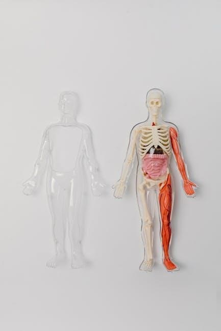
The Skeletal System
This section focuses on the examination and identification of skeletal structures‚ including major bones and landmarks. It explores the relationship between skeletal anatomy and functional movements‚ preparing students to articulate joints and analyze their mechanics through hands-on activities.
2.1 Preparation and Examination of Skeletal Specimens
Proper preparation involves cleaning and preserving skeletal specimens to ensure safe handling and detailed observation. Students learn to identify and examine bones‚ focusing on structural features and anatomical landmarks. This step is crucial for understanding skeletal anatomy and its role in movement and support. Gloves and goggles are essential for safety during the process.
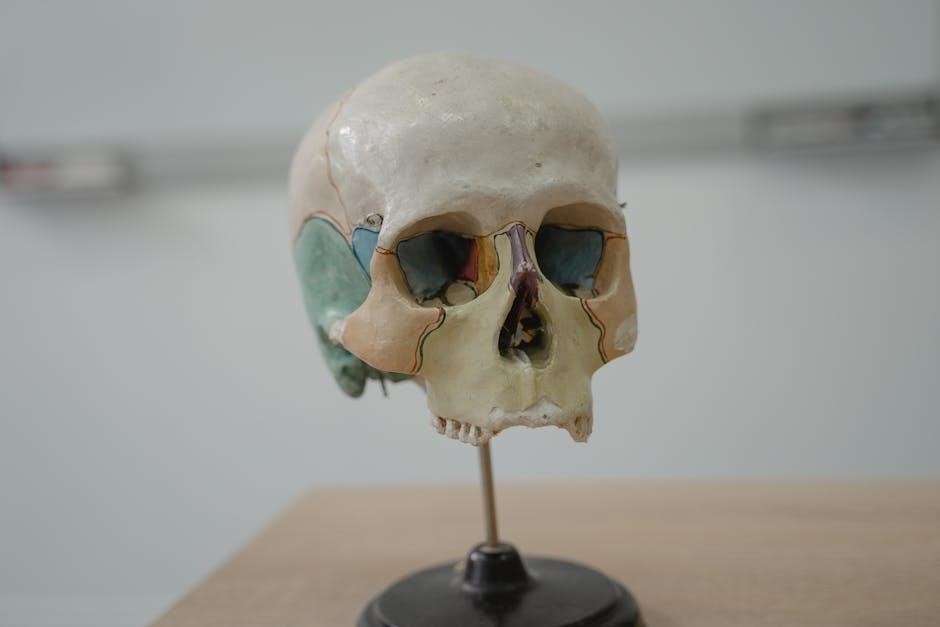
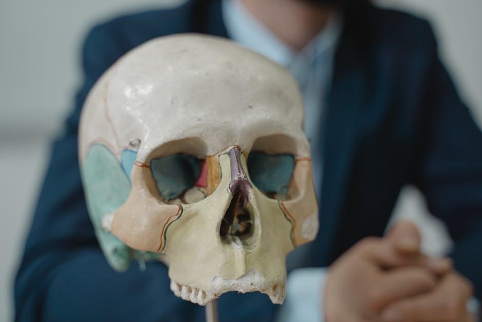
2.2 Identification of Major Bones and Landmarks
This section focuses on recognizing key bones and their anatomical landmarks. Students learn to identify major bones like the femur‚ humerus‚ and cranium‚ along with significant features such as the acetabulum‚ condyles‚ and mandible. Using detailed diagrams and 3D models‚ participants gain hands-on experience in distinguishing bone structures‚ enhancing their understanding of skeletal anatomy and its functional significance in the human body.
2.3 Hands-On Activity: Articulating Joints
This activity involves assembling and disassembling joint models to understand their structure and function. Students observe how bones articulate and classify joint types based on movement. Interactive exercises include simulating joint dislocations and analyzing the role of ligaments and cartilage. This practical approach helps visualize joint mechanics‚ reinforcing theoretical concepts and improving anatomical understanding through tactile learning experiences.
The Muscular System
This section explores the structure and function of muscles‚ emphasizing voluntary and involuntary muscle types. It includes dissection of major muscle groups and practical exercises for understanding movement mechanics.
3.1 Dissection and Exploration of Major Muscle Groups
This section provides hands-on experience with dissecting major muscle groups‚ allowing students to identify and examine muscle structures‚ origins‚ and insertions. Through careful dissection‚ students gain insight into muscle arrangement and function‚ bridging lecture concepts with practical observation. Emphasis is placed on safety‚ proper technique‚ and ethical specimen handling to ensure a meaningful learning experience.
3.2 Understanding Muscle Function and Movement
This section explores the mechanisms of muscle function‚ focusing on contraction types‚ such as isotonic and isometric movements. Students learn how muscles interact with bones and joints to produce motion. Practical exercises‚ including palpation‚ help identify muscle actions in real-time. This hands-on approach clarifies how muscles contribute to posture‚ locomotion‚ and overall bodily movements‚ reinforcing theoretical concepts with observable outcomes.
3.3 Practical Exercise: Palpation of Muscles
This exercise teaches students to identify and assess muscle structures through palpation. Using anatomical charts and guided instruction‚ participants locate key muscles‚ observe their texture‚ and understand their movements. This hands-on activity enhances tactile awareness and familiarity with muscle anatomy‚ preparing students for clinical applications and improving their diagnostic skills in real-world scenarios.
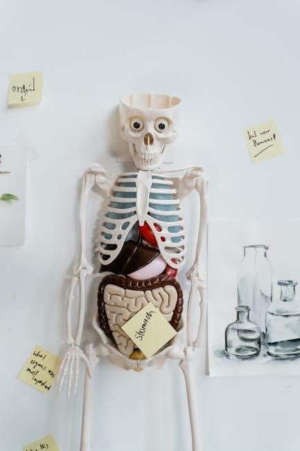
The Digestive System
This section explores the structure and function of the digestive system through external and internal examinations of organs‚ enzyme analysis‚ and comparative species studies to enhance understanding of digestion processes.
4.1 External and Internal Examination of Digestive Organs
This section involves the macroscopic and microscopic examination of digestive organs‚ focusing on their structure and anatomical landmarks. Students will handle specimens to identify external features and internal chambers‚ such as the stomach’s mucosa or intestinal villi. The use of dissecting scopes and magnification tools aids in detailed observation‚ correlating findings with textbook descriptions to enhance understanding of digestive anatomy and function.
4.2 Laboratory Investigation of Digestive Enzymes
This section focuses on analyzing the activity of digestive enzymes‚ such as amylase‚ lipase‚ and trypsin‚ through controlled experiments. Students measure enzyme effectiveness under varying conditions‚ including pH levels and inhibitor presence. Practical exercises involve simulating digestion processes and observing biochemical reactions. These investigations highlight enzyme specificity and their critical role in nutrient breakdown‚ aligning theoretical knowledge with experimental outcomes to deepen understanding of digestive biochemistry.
4.3 Activity: Comparing Digestive Processes in Different Species
This activity involves comparing the digestive systems of various species‚ such as herbivores‚ carnivores‚ and omnivores. Students analyze anatomical structures‚ like stomachs and intestines‚ and observe physiological differences in enzyme production. Practical exercises include simulating digestion in different species and discussing adaptations related to diet. This comparative approach highlights evolutionary specializations and their impact on nutrient absorption and energy utilization.
The Circulatory System
This section explores the structure and function of the circulatory system‚ focusing on blood composition‚ heart anatomy‚ and vascular dynamics. Hands-on activities include blood typing‚ heart dissection‚ and blood pressure measurement to understand cardiovascular health and regulation.
5.1 Blood Typing and Hematological Analysis
This section introduces blood typing‚ focusing on ABO and Rh factor identification. Students learn hematological analysis‚ including blood cell counting and smear preparation. Practical exercises emphasize understanding blood composition‚ clotting mechanisms‚ and the importance of safety protocols when handling biological specimens. These activities provide insights into blood disorders and diagnostic techniques used in clinical settings.
5.2 Dissection of Blood Vessels and Hearts
This section focuses on the detailed dissection of blood vessels and hearts to examine their structural anatomy. Students identify key features such as chambers‚ valves‚ and vascular branching patterns. Hands-on dissection enhances understanding of circulatory pathways and cardiac function. Emphasis is placed on proper dissection techniques‚ specimen handling‚ and correlating anatomical structures with their physiological roles in blood circulation and overall health.
5.3 Measurement of Blood Pressure and Heart Rate
This section introduces techniques for measuring blood pressure and heart rate‚ essential for assessing cardiovascular health. Students learn to use sphygmomanometers and stethoscopes to record systolic and diastolic pressures. Heart rate is measured through pulse palpation or ECG simulations. Emphasis is placed on understanding normal ranges‚ factors influencing measurements‚ and the importance of accurate data recording for diagnosing physiological conditions and monitoring health trends.
The Respiratory System
This section explores the respiratory system’s structure and function through detailed examinations‚ hands-on activities‚ and experiments‚ providing practical insights into breathing mechanisms and gas exchange.
6.1 Examination of Lung Structure and Function
This section focuses on the gross anatomy of lungs‚ including lobes‚ pleura‚ and airways. Students examine histological slides to identify alveoli and bronchioles. Functional assessments include spirometry to measure lung capacity and gas exchange demonstrations. Practical exercises reinforce understanding of breathing mechanisms‚ ventilation‚ and oxygenation‚ correlating structure with physiological processes.
6.2 Spirometry and Breathing Exercises
Students learn to measure lung function using spirometry‚ assessing vital capacity‚ tidal volume‚ and forced expiratory rate. Breathing exercises‚ such as diaphragmatic breathing‚ are practiced to enhance lung efficiency. Data interpretation and comparison with normal values help understand respiratory health. These activities emphasize the role of breathing techniques in improving pulmonary function and overall well-being‚ while reinforcing physiological concepts.
6.3 Laboratory Demonstration of Gas Exchange
This section demonstrates the process of gas exchange in the lungs‚ focusing on oxygen and carbon dioxide diffusion across alveolar membranes. Students observe how oxygen binds with hemoglobin and CO2 is transported for exhalation. The importance of concentration gradients and proper ventilation is highlighted. Practical exercises using lung models simulate gas exchange‚ reinforcing its critical role in respiration and overall bodily function.
The Nervous System
This section explores the structure and function of the nervous system‚ focusing on neurons‚ reflexes‚ and sensory responses. Practical exercises include identifying cranial nerves and analyzing nerve function.
7.1 Exploration of Neuron Structure and Function
This section focuses on understanding the structure and function of neurons‚ including dendrites‚ axons‚ and synapses. Hands-on activities involve examining neuron models‚ staining nerve tissue samples‚ and observing nerve impulses. Students learn how neurons transmit signals and communicate through synapses‚ essential for nervous system function. These exercises provide insights into neural communication mechanisms and their role in human physiology.
7.2 Reflex Testing and Sensory Response Analysis
This section covers the assessment of reflexes‚ including deep tendon and superficial reflexes‚ to evaluate nervous system function. Students participate in testing patellar‚ Achilles‚ and plantar reflexes. Sensory response analysis involves examining reactions to stimuli like touch‚ pain‚ and vibration. Hands-on activities include documenting responses and interpreting results to understand neural control mechanisms and sensory pathways in human physiology.
7.3 Hands-On Activity: Identifying Cranial Nerves
This activity focuses on the identification and localization of the 12 cranial nerves through dissection and observation. Students will trace nerve pathways‚ explore their functions‚ and test reflexes associated with specific nerves‚ such as the optic‚ facial‚ and vagus nerves. Practical exercises include nerve stimulation and sensory testing‚ using anatomical charts and cadaveric specimens to reinforce understanding of cranial nerve roles in sensory and motor functions.
Histology and Microscopy
This section introduces histology and microscopy‚ essential tools for studying tissue and cellular structures. Students learn to prepare and examine samples‚ gaining skills in identifying tissue types and understanding cellular functions through microscopic analysis.
8.1 Preparing and Staining Tissue Samples
Preparing and staining tissue samples involves fixation‚ dehydration‚ sectioning‚ and staining to preserve and enhance cellular structures. Fixation stabilizes tissues‚ while dehydration prepares them for embedding. Sectioning creates thin slices for microscopy; Staining‚ such as hematoxylin and eosin (H&E)‚ highlights cellular details. Proper techniques ensure clear visualization of tissue architecture and cellular components‚ aiding in accurate histological analysis and diagnosis.
8.2 Microscopic Examination of Epithelial‚ Connective‚ and Muscle Tissues
Microscopic examination allows detailed study of tissue structures. Epithelial tissues show tight junctions and layered arrangements‚ while connective tissues reveal a matrix with embedded cells like fibroblasts and collagen fibers. Muscle tissues exhibit striated patterns due to sarcomeres. Proper staining enhances visibility of these features‚ enabling identification of tissue types and their specialized functions under magnification.
8.4 Activity: Identifying Cellular Structures Under a Microscope
Under the microscope‚ students examine prepared slides of epithelial‚ connective‚ and muscle tissues. Proper focus and illumination techniques are demonstrated to enhance visibility of cellular details. The activity involves identifying key structures such as cell membranes‚ nuclei‚ and cytoplasm‚ as well as specialized features like keratin in epithelial cells or sarcomeres in muscle fibers. This hands-on exercise reinforces microscopic observation skills and tissue identification.
Integration of Laboratory Experiences
This section integrates laboratory findings with lecture material‚ offering case studies and a final project for comprehensive understanding and application‚ enhancing learning outcomes.
9.1 Correlating Laboratory Findings with Lecture Material
This section emphasizes linking hands-on laboratory experiments with theoretical concepts discussed in lectures. By analyzing lab results and connecting them to classroom teachings‚ students deepen their understanding of anatomical and physiological principles. This integration enhances critical thinking and reinforces key concepts‚ preparing learners for practical applications in healthcare and research.
9.2 Case Studies in Anatomy and Physiology
Case studies provide real-world applications of anatomical and physiological concepts‚ allowing students to analyze and interpret clinical scenarios. Through detailed examinations of medical conditions‚ injuries‚ or structural anomalies‚ learners develop problem-solving skills and apply theoretical knowledge to practical situations. These studies enhance understanding of human body functions and prepare students for diagnosing and treating disorders in clinical settings.
9.3 Final Project: Comprehensive Lab Report and Presentation
The final project requires students to synthesize knowledge from the laboratory manual by preparing a detailed report and presentation. The report includes experimental data‚ observations‚ and conclusions‚ while the presentation communicates findings effectively. This project fosters scientific communication skills‚ critical thinking‚ and the ability to articulate complex anatomical and physiological concepts clearly. Peer feedback and evaluation refine presentation techniques and understanding;